You probably have a mental snapshot of a cell from eighth-grade biology. It’s that neon-green brick or the pinkish, jelly-like blob floating in a textbook. Honestly, those diagrams are a bit of a lie. They’re "idealized." In reality, when you look at a picture of plant cell and animal cell structures under a high-powered electron microscope, things get messy, crowded, and incredibly kinetic. They aren't static little rooms; they are chaotic chemical cities.
Cells are the literal hardware of life. If you’re trying to understand why your muscles ache after a workout or how a sunflower tracks the sun, the answer is buried in the architecture of these microscopic units. Most people think the only difference is that plants have a "wall" and animals don't. That’s like saying the only difference between a tank and a convertible is the paint job. It goes way deeper into how energy is handled and how these organisms survive.
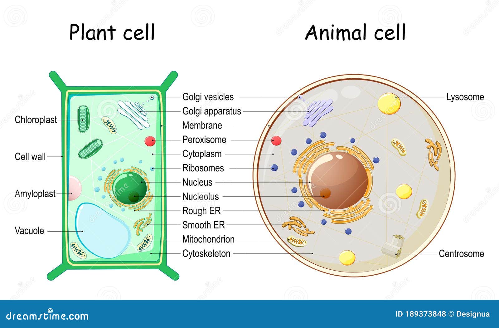
The Big Differences in a Picture of Plant Cell and Animal Cell
Let’s get the obvious stuff out of the way first. When you see a picture of plant cell and animal cell side-by-side, the plant cell usually looks like a sturdy rectangle. That’s the cell wall. It’s made of cellulose—the same stuff in your cotton t-shirt. Animal cells? They’re floppy. They have a plasma membrane, but without that rigid outer skeleton, they can take on all sorts of weird shapes, like the long, spindly reach of a neuron or the smooth disc of a red blood cell.
The Solar Panels vs. The Power Plants
Plants are basically solar-powered. Inside a plant cell, you’ll see these green ovals called chloroplasts. They contain chlorophyll, which captures light. Animal cells don't have these because, well, we eat our energy instead of soaking it up from the sky. However, both cells have mitochondria. You’ve heard the meme: the powerhouse of the cell. It’s true. Mitochondria take glucose and turn it into ATP ($C_{10}H_{16}N_{5}O_{13}P_{3}$), the universal currency of biological energy.
Storage Wars: The Vacuole
Look at a plant cell under a microscope. You’ll notice a massive "bubble" in the middle. That’s the central vacuole. It’s huge. It stores water and creates "turgor pressure." When you forget to water your fiddle-leaf fig and it wilts, it’s because those vacuoles have deflated. Animal cells have vacuoles too, but they’re tiny, temporary, and usually used for getting rid of waste or moving things around. They aren't the structural pillars that they are in the plant world.
Why the Shapes Actually Matter
Form follows function. Always.
Because plants can’t run away from a predator or move to a shady spot, they need to be tough. The cell wall provides that. It allows trees to grow hundreds of feet tall without a skeleton. On the flip side, animal cells need flexibility. Think about your white blood cells. They have to squeeze through tiny capillary gaps to chase down bacteria. If they had a rigid cell wall, you’d be dead. Your immune system would be stuck in traffic.
The "fluid mosaic model" is a term scientists use to describe the animal cell membrane. It’s not a solid skin; it’s more like a sea of lipids with proteins floating in it like icebergs. This allows the cell to "swallow" things (endocytosis) or "spit" things out (exocytosis) with ease. Plants do this too, but they have to work around that cellulose fence.
The Secret Organelles Nobody Mentions
Everyone talks about the nucleus. Sure, it’s the brain. It holds the DNA. But have you looked at the Endoplasmic Reticulum (ER)? In a picture of plant cell and animal cell, the ER looks like a bunch of folded ribbons tucked right next to the nucleus.
There’s "Rough" ER and "Smooth" ER. The rough stuff is covered in ribosomes, making it look like it’s been rolled in sand. This is the protein factory. The smooth ER is where lipids are made and toxins are neutralized. If you drink a glass of wine, the smooth ER in your liver cells goes into overdrive to process the alcohol.
Then there's the Golgi apparatus. Think of it as the FedEx of the cell. It takes the proteins made by the ER, packages them into little bubbles called vesicles, and tags them with a "zip code" so they know where to go in the body. Without the Golgi, your cells would just be piles of useless raw materials.
Centrioles and Lysosomes
Here is a nuance that often gets skipped: centrioles. Usually, you’ll only see these in an animal cell picture. They look like little bundles of pasta and help the cell divide. Most plants don't have them; they use a different method to organize their DNA during division.
And lysosomes? They’re the "suicide bags" or garbage disposals. They are packed with enzymes that break down waste. While plants have similar structures in their vacuoles, the animal lysosome is a specialized, highly acidic little sphere dedicated to cleaning up the cellular neighborhood.
Misconceptions That Mess With Your Head
One of the biggest mistakes people make when looking at a picture of plant cell and animal cell is thinking they are two-dimensional. They aren't circles and squares. They are 3D globes and polyhedrons.
Another weird one? The idea that animal cells are "simple." They’re actually incredibly complex because they have to coordinate with nervous systems, muscle contractions, and hormonal signals. A plant cell is a marvel of self-sufficiency, but an animal cell is a marvel of communication.
Also, don't think that because plants have cell walls, they are "locked" away. They have these tiny tunnels called plasmodesmata. These are literal holes in the cell walls that let neighbor cells swap soup, signals, and nutrients. It’s a literal network.
The Real-World Impact: Why Should You Care?
Understanding these differences isn't just for passing a test. It’s how we create medicine.
Antibiotics like penicillin work by attacking the way bacteria build their cell walls. Since your human cells don't have cell walls, the medicine kills the bacteria but leaves you alone. If our cells were structured like plant cells, penicillin would be a poison, not a cure.
In agriculture, scientists look at chloroplast efficiency to try and grow crops that need less water or produce more food. Every time you eat a salad or take an aspirin (which originally came from willow bark), you’re interacting with the specific chemistry of these cell types.
How to Correctly Identify These Cells Under a Microscope
If you ever find yourself looking through a lens, here is how you tell them apart without a label:
- Look for the Border: Is it thick and dark? Plant. Is it faint and irregular? Animal.
- Check the Color: See green spots? Chloroplasts mean it's a plant (usually).
- The Nucleus Position: In a plant cell, the big vacuole often pushes the nucleus to the side. In an animal cell, the nucleus is usually hanging out near the center.
- Staining: Scientists often use Methylene Blue for animal cells (like your cheek cells) because it makes the nucleus pop. For plants, Iodine is common because it reacts with the starch stored in the cell.
Cells are the frontier. We are still discovering new things about how the "cytoskeleton"—a network of protein fibers—actually moves things around inside. It’s not just floating goop; it’s a highly organized lattice that acts like a conveyor belt.
Moving Toward a Better Understanding
To really grasp this, don't just stare at one picture of plant cell and animal cell. Look at diverse versions. Look at specialized cells. Compare a leaf cell to a root cell; the root cell won't have chloroplasts because it lives in the dark. Compare a skin cell to a heart muscle cell; the heart cell will be packed with way more mitochondria because it never gets to stop beating.
Next Steps for Deepening Your Knowledge:
- Search for 3D Cell Models: Use interactive tools like the "Cell Alive" website to rotate the cells. It changes your perspective on how crowded the cytoplasm really is.
- Observe Real Cells: If you can get a cheap 400x microscope, scrape some onion skin (plant) and some of your own cheek cells (animal). Use a drop of food coloring if you don't have professional stains.
- Study the Cytoskeleton: Research "microtubules" and "actin filaments." This is the "hidden" infrastructure that standard diagrams often leave out to keep things simple.
- Explore Extremophiles: Look up how cells differ in organisms that live in volcanoes or at the bottom of the ocean. It’ll blow your mind how much the "standard" cell model can be pushed.
Understanding the blueprint of life starts with these two basic units. Once you see the complexity, a simple blade of grass or the skin on your hand never looks the same again.